Hi All, We have the latest MG modality from Hologic (Hologic 3Dimension) which was installed at the site very recently. We have Philips Vue PACS ver: 12.2.5.
We are one of the largest Cancer care and referral center in the Middle east region. Hence for majority of the mammogram studies performed at center we do the tomosynthesis as well. The image size of the tomosynthesis studies are ranging from a minimum of 6 GB to 18 GB. This consuming a lot of space in our PACS storage and even challenging as these studies wont fit into a CD or DVD. Vendor states that the huge size is due to the high definition images that are acquired during 3D (DBT). …
Anyone has faced the similar issue with Hologic 3dimension DBT image storage… Any insights on what can be done to reduce the DBT study size? Please share any ideas/feedback in this regard.
Forum Colleagues,
Any feedback/suggestions on the below mentioned scenario will be of great help…
"Hi All, We have the latest MG modality from Hologic (Hologic 3Dimension) which was installed at the site very recently. We have Philips Vue PACS ver: 12.2.5.
We are one of the largest Cancer care and referral center in the Middle east region. Hence for majority of the mammogram studies performed at center we do the tomosynthesis as well. The image size of the tomosynthesis studies are ranging from a minimum of 6 GB to 18 GB. This consuming a lot of space in our PACS storage and even challenging as these studies wont fit into a CD or DVD. Vendor states that the huge size is due to the high definition images that are acquired during 3D (DBT). …
Anyone has faced the similar issue with Hologic 3dimension DBT image storage… Any insights on what can be done to reduce the DBT study size? Please share any ideas/feedback in this regard."
It sounds like you might not be using any compression - it would be normal to use JPEG2000 lossless compression on these objects (and all DICOM really!), which reduces the size as stored substantially.
DICOM can store images in different encodings, some with lower storage requirements than others, however, without access to the specific hardware, it’s difficult to make any helpful suggestions.
That image size does seem very large. From the lack of responses so far, I doubt anyone can (quickly) solve your problem. I would escalate with the vendor.
Thanks for the responses Gentlemen,
I had already suggested the vendor to check on the image compression techniques and they did a configuration change in the machine as well however it was of no luck… still we have high image sizes for DBT’s. They have escalated the matter internally and we are yet awaiting a response back from them. Vendor is claiming that huge DBT image size is due to the HD images that are acquired by the latest 3 dimension machine from Hologic.
It’s a bit of a long-shot but you could try to download one of the DICOMs and have a closer look at what is being sent e.g. using the dcmdump utility (from dcmtk). This will give you a better idea of what is being sent from the machine and you might find some opportunities for optimisation.
We use Hologic machines, but I’m not sure which ones. Just as a reference, one of our Tomo studies is circa 2GB without compression and 0.55GB with compression. I could well believe some of it is to do with the Vendor’s set up, especially if they are using high definition acquisitions, with my Radiographer hat on, I’d want to know what technology they were using to get high definition images without high levels of irradiation.
There are various answers to this to be considered.
On a macro scale, what are you going to store to PACS. Just the tomo reconstructions or tomo + raw data. Probably just the tomo unless you have a medico legal reason to store raw too. That already cuts down the quantity of stored data.
If you don’t want to reduce the quantity of pixels you are storing, you could address the issue by increasing your PACS storage (a contractual answer rather than a technical answer - and often the application of a technical solution has a cost consequence as suppliers change config - you should probably explore both).
Use of compression. Most countries insist on lossless compression (not losing data). DICOM supports JPEG 2000 lossless pixel representation - for mammo images this can give around 3 or 3.5:1 compression - but I am not sure about the tomosynthesis image sets - there may be residual pixel fluctuations from the recon algorithm n the spaces we see as zeros (low level noise) and this will reduce the level of compression that is achievable - and possibly varies between tomosynthesis algorithms (from manufacturer to manufacturer). Interesting physics study here just by inspecting compression ratios between suppliers.
Anyway - they are still going to be large studies even with compression - they will need planning for.
Forgot to mention - where to apply the compression. The modality may have an option to export lossless comresed images but also the PACS may be able to convert to lossless compressed on ingestion.
Hi Robin, Thanks for your reply. Addressing the point number 1 in your message, when I noticed this behavior first I too had the feeling that modality is sending RAW images however I have the confirmation from Hologic service team that modality isn’t sending the RAW data. It is just sending the HD DBT images.
Also I have checked with the Mammographers and I understand that the number of images totally depends on the breast thickness & various other patient conditions…
We just started the center and PACS is a new installation so we have got a lot of storage at this moment. Having said that, we haven’t considered such huge sizes for MG DBT images while building the PACS storage for our site. Since we are just starting storage wont be an issue but within a year or so we will facing issues with storage as well… Also I am afraid of network congestion/traffic that can be caused due to this high image size / study and the number of MG exams that we perform a day is also high…
I will discuss with the vendor on the JPEG 2000 lossless compression technique as well…
You need to make sure that JPEG2000 is the first option on the list of transfer syntaxes on the Hologic, and that the PACS is configured to accept JPEG2000 transfer syntax (and preferably has it high on their ordering too). This will help with the network traffic and transfer speeds.
The projection data isn’t really important as the file sizes are small compared to the tomosynthesis.
The next thing is then to configure the PACS to store images as JPEG2000 compression, to reduce the storage requirement for each exam.
And yes, the size of the DBT images are almost directly proportional to the compressed breast thickness.
Hi Robin,
I have configured PACS for JPEG 2000 lossless transfer syntax and Hologic vendor also confirmed that they have configured the machine for same transfer syntax. However the I haven’t seen any reduction in the MG DBT study sizes after the changes also.
Model of Hologic machine: 3 Dimensions - 3D with clarity HD
I have configured PACS for JPEG 2000 lossless transfer syntax and Hologic vendor also confirmed that they have configured the machine for same transfer syntax. However the I haven’t seen any reduction in the MG DBT study sizes after the changes also.
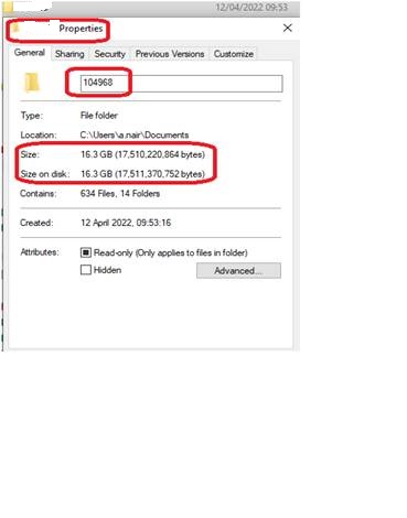
I am attaching a file size of a MG study that was sent to Vue PACS from Hologic 3dimension. As you can see its 16.3 GB. .we have a lot of similar kind of studies. Vendors have assured that they aren’t sending RAW data along with the DBT & 2D images. Also I have applied the JPEG 2000 lossless transfer syntax as well…
We have MG model - Hologic 3 dimension - (3D with HD clarity). Regarding the HD images - Vendor explains that the HD clarity images can be acquired due to the pixel size is 70 microns of the machine detector. Radiation dose remains low as it is in other Hologic MG machines.
If you have exported the study from the PACS to your local machine, it is possible that it has been transcoded again to not be compressed anymore.
However, in the screenshot below you are showing a collection of 634 files – how many of them are the DICOM images? I’m guessing that that includes all the DICOM viewing software that often gets written on export.
We too have Hologic 3Dimensions systems, with the 70 µm reconstructed pixel size in tomosynthesis mode. Uncompressed, the typical image sizes range from <800 MB for 2 cm, up to 2.6 GB for 7 cm reconstructions (from our QA images). You can add about 300 MB for the projection images if you store those to PACS, and about 25 MB for the synthetic 2D image if you create that.
If you had a large lady with four views you might get to 10 GB, uncompressed, but most would be much less than that, and you’d want to store them compressed too.
Is your PACS vendor able to tell you how much space they take stored on their system?